ORBITAL-FACIAL BONE FRACTURES:
The ocular globe (eyeball) is housed and protected within the bony orbits of the facial bones. These orbits are made up of 8 individual and distinct bones. When a patient sustains a blunt traumatic injury to the orbital region it is common that one or multiple of these bones will fracture. Common injuries which result in orbital bone fractures include: Sports related injuries such as baseball, or tennis in which one might get directly hit in the eye with a ball. Traumatic injuries of the orbital region such as involving a motor vehicle collision, work related injury or “punch” to the orbital region. In all of these cases a CT scan of the face and orbital bones is required as part of the overall medical workup. It is also recommended that a complete ophthalmology exam be performed to evaluate and rule out any injury to the ocular globe.
If the diagnosis of an orbital bone fracture is confirmed either on physical exam and or CT scan imaging, then Dr. Praveen will review your options either for surgical intervention or close observation of the injury. If surgical intervention is recommended Dr. Praveen will review the procedure of choice, depending on your individual case for reconstruction of your orbital facial bone fractures.
ZYGOMA (CHEEK BONE-TRIPOD) FACIAL FRACTURES:
The zygoma is commonly known as the cheek bone. It anatomically lies between the orbital bone above it and the upper jaw (maxilla) below. It is similarly commonly fractured by sustaining a direct or indirect trauma or blow to this portion of the face. In the past the misnomer of “Tripod Fracture” has been assigned to this type of fracture. Once again a CT scan of the facial bones along with a complete physical exam is required to evaluate and diagnose the facial bone fractures accurately. If the diagnosis of a zygoma fracture is confirmed, in most cases surgery is required to reposition the facial bone fracture and fixate it to the surrounding stable bones with micro plates and screws.
MAXILLA AND MANDIBLE (UPPER AND LOWER JAWS) FACIAL BONE FRACTURES:
The maxilla (upper jaw) and mandible (lower jaw) are the “teeth bearing” bones of the face. These are the facial bones in which both the primary (baby teeth) and permanent teeth are formed and located. Fractures of these bones similarly occur from either direct or indirect blunt force to the facial bones. Fracture of these facial bones may also be associated with fractures of the teeth or tooth roots within the region of the bony fracture sites. Upon evaluation of a patient with a fracture of the maxilla or mandible it is commonly noted and the patient reports that the “teeth do not fit together” correctly. The patient now has a mal-occlusion due to the fact that several teeth, in either the maxilla or mandible are now contacting the opposing jaw in a different way than they did prior to the accident.
Treatment of fractures of the maxilla and or mandible are most commonly performed with surgical re-alignment of the fractured facial bones and fixating the fractures with micro plates and screws. It is often required that metal braces be placed on the upper and lower teeth prior to the placement of the micro plates and screws. These braces, known as Arch Bars, are placed in the operating room and usually removed at the end of the case. In some cases where the fractured bones are unstable, even after the placement of the plates and screw, the arch bars may remain in place for up to six weeks after the surgical fixation. This is to provide additional support to the alignment of the bony fractures during the healing phase. If there are any broken teeth or tooth roots, these may need to be removed either at the time of the facial bone fracture surgery or afterwards.
FOREHEAD, FRONTAL BONE AND FRONTAL SINUSES
The bony support of the forehead is made up of the Frontal Bones. Within the frontal bones in the region above the nasal bridge (glabella) lies the frontal sinuses. In cases where the forehead sustains blunt trauma the frontal sinuses are often fractured. The frontal sinuses have both a front (anterior) wall and a back (posterior) wall. In cases where both the front and back wall are fractured, this often requires the assistance of a neurosurgeon to assist in the reconstruction of the frontal sinus fracture. In cases in which only the front wall of the frontal sinus is fractured, Dr. Praveen routinely reconstructs this region with micro plates, screws and often bone grafts. As in the above cases, a complete CT evaluation of the involved region is required prior to treatment. The bony fractured segments are realigned and fixated with micro plates and screws. Fractures of the frontal sinuses may require long term issues which need to be addresses if the drainage system of the sinuses (nasal-frontal ducts) is damaged due to the trauma of the frontal bone fractures.
NOSE AND NASAL BONES
Nasal bone fractures are the most common facial bone injured. Traumatic nasal bone injuries, besides resulting in a crooked nose may result in other issues. Nasal airway obstruction secondary to nasal septal deviation may result. In most cases, as long as the patient has no other emergent medical issues, it is usually recommended to treat nasal bone fractures if the nasal bones are displaced or if they are causing nasal airway obstruction. Even in cases where the nasal bones and or nasal septum are aligned, straightened and reduced, there is always the possibility that due to normal healing and scar tissue, further surgery may be required in the future.
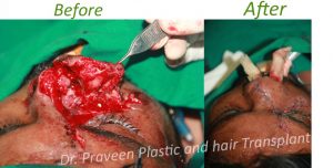
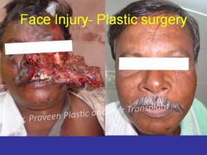
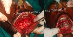
FACE TRAUMA